New method offers hope of fewer fractures
Around one thousand Swedes could be spared from a hip fracture each year if a new method to identify the risk of osteoporotic fractures were to be introduced in healthcare. This is the view of the researchers at Lund University’s Faculty of Engineering (LTH) who are behind a new 3D-simulation method. The results were recently published in the Journal of Bone and Mineral Research.
Jessika Sellergren – Published 13 September 2023
Osteoporosis causes 120,000 bone fractures in Sweden every year – including 18,000 hip fractures – and half of all those affected will sustain more fractures. The condition entails a reduction in bone density, a weakening of bone structure and an increased risk of fractures. Osteoporosis mainly affects the elderly.
Current method misses around half of those at high risk – but new method can catch thousands more every year
The current diagnosis technique for osteoporosis is a bone density measurement – a 2D X-ray examination that in general is first used once the patient has already sustained a fracture.
However, measurement of the skeleton’s bone density does not detect all those with a high risk of a fracture and around half of the people in the risk zone are missed, points out Hanna Isaksson, professor of biomedical engineering at Lund University’s Faculty of Engineering (LTH).
“Up to now there have been no reliable methods for identifying the condition at an early stage, importantly before the first fracture occurs,” she says.
A greater percentage of those with a high risk of sustaining a hip fracture could be identified using a new calculation and simulation method that Hanna Isaksson and her research colleagues have developed. This was shown in the results of a study that evaluated the method based on patient data from 2,000 people – the results were recently published in the Journal of Bone and Mineral Research.
“Using the new model, we could identify around 1,000 more people per year with a high risk of hip fracture. The results suggest that the method can also be used to identify osteoporosis before the first fracture occurs,” says Hanna Isaksson.
When 2D becomes 3D, more high-risk patients can be identified
Lorenzo Grassi, associate senior lecturer in biomedical engineering at Lund University, was responsible for evaluating the method. He explains that the new method uses 2D X-ray images from bone density measurements to produce 3D models of the thigh bone.
“The shift from 2D to 3D is conducted with the help of a computer-simulated template that describes how the bone’s geometry and bone density varies in the population.”
The 3D model of the thigh bone enables the simulation of different situations and scenarios that may have an effect, for example in the case of a fall – information that makes it easier to assess the risk of fractures.
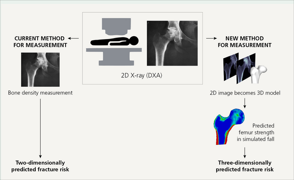
In current diagnostics, bone density measurements from 2D X-ray images are used to estimate the risk of fracture. The new method creates 3D models from 2D images to calculate thigh bone strength as a basis for estimating the risk of fracture. Illustration: Lorenzo Grassi
Weakness in the thigh bone is a risk factor
Lorenzo Grassi has conducted simulations on data from over 400 people who previously underwent X-ray-based bone density measurements. Of these, some sustained a hip fracture within a ten-year period after the examination, while others did not. When the measurement results from the two methods were compared, it was clear that the 3D method is better at predicting the risk of a hip fracture than the bone density measurement.
“If the people who actually sustained a hip fracture had been examined using the new method, those with low bone strength could have been identified and some of the bone fractures could have been avoided,” says Hanna Isaksson.
Lorenzo Grassi explains that the simulations are important parts of the puzzle in understanding the factors that affect the risk of sustaining a fracture:
“As a person can fall in many different ways, the person can also be injured in various ways. Using the new method, it is possible to simulate the different ways in which a person can fall – information that enables us to calculate the force required to fracture a bone.”
It is also possible to generate specific values for the bone strength of an individual’s skeleton.
“This is particularly important as a weak skeleton is one of the biggest risk factors for osteoporosis-related fractures,” says Lorenzo Grassi.
Enhanced quality of life and lower costs
Looking ahead, the researchers are planning more evaluations of the measurement method that they believe can be an important supplement to today’s diagnostics. They hope that the method will be integrated in care relating to osteoporosis, in both healthcare centres and specialist healthcare.
The earlier detection of osteoporosis can help to improve quality of life for patients and reduce costs for society,” explains Hanna Isaksson.
“The annual cost that arises due to osteoporosis in Sweden was estimated for 2019 at SEK 23 billion, a cost that increases every year. This trend needs to be reversed.”
More accurate diagnosis could also reduce the number of deaths.
“Of those who sustain a hip fracture, 24 per cent die within one year,” she says.
Hanna Isaksson
Professor of Biomedical Engineering at the Faculty of Engineering (LTH)
Assistant Coordinator of the LU Profile Area: Proactive Ageing
Lorenzo Grassi
Associate Senior Lecturer in Biomedical Engineering at the Faculty of Engineering (LTH)
Testing the method
The researchers in biomedical engineering at Lund University’s Faculty of Engineering (LTH), together with clinicians in Malmö and Uppsala, tested the measurement method using data from 2,000 patients aged between 69 and 80 who participated in the ten-year study MrOS – The Osteoporotic Fractures in Men Study. Over 400 simulations have been conducted based on the patient data. The researchers will also test the method using patient data from women.
“The technology behind the method was developed based on information from both women and men, and we expect that the method works just as well in identifying those people, regardless of gender, with a high risk of hip fractures. We are conducting a computer simulation study at the moment based on a clinical study of the risk of fracture among women,” says Hanna Isaksson.
Read more in the Journal of Bone and Mineral Research
An article on the measurement method has been published in the Journal of Bone and Mineral Research: 3D finite element models reconstructed from 2D DXA images improve hip fracture prediction compared to areas BMD in MROS Sweden cohort
The authors of the article are:
- Lorenzo Grassi, Biomedical Engineering at LTH, Lund University
- Hanna Isaksson, Biomedical Engineering at LTH, Lund University
- Magnus K. Karlsson, Orthopaedics, Lund University
- Björn E. Rosengren, Orthopaedics, Lund University
- Lars Jehpsson, Orthopaedics, Lund University
- Sami P. Väänänen, University of Eastern Finland
- Östen Ljunggren, Uppsala University